Prelude: A Quiet Shift from Clay to Light
The clinic used to hum like an old amp: impressions, plaster, the patient shifting in the booth. Then the first clear note arrived — a Photogrammetry 3D scanner — and the tone changed. This is an evolution, not a flip of a switch: a series of small, deliberate moves that reshape diagnosis, planning, and appliances. The story that follows traces those moves in lived detail, the crescendos and the missed beats, from first capture to final deliverable.
First Measures: Why Capture Matters
Capture is the opening riff. A precise scan gives you pitch-perfect models; a sloppy capture leaves you tuning forever. Clinicians I’ve worked with report that consistent lighting, stable markers, and controlled patient movement cut re-scans in half. Do three quick checks before you stop: surface completeness, occlusal contact, and soft-tissue margins. If any of those sound off, rescan immediately. Small corrections now save wasted lab nights later.
The Rhythm of Tools: Where Photogrammetry Fits
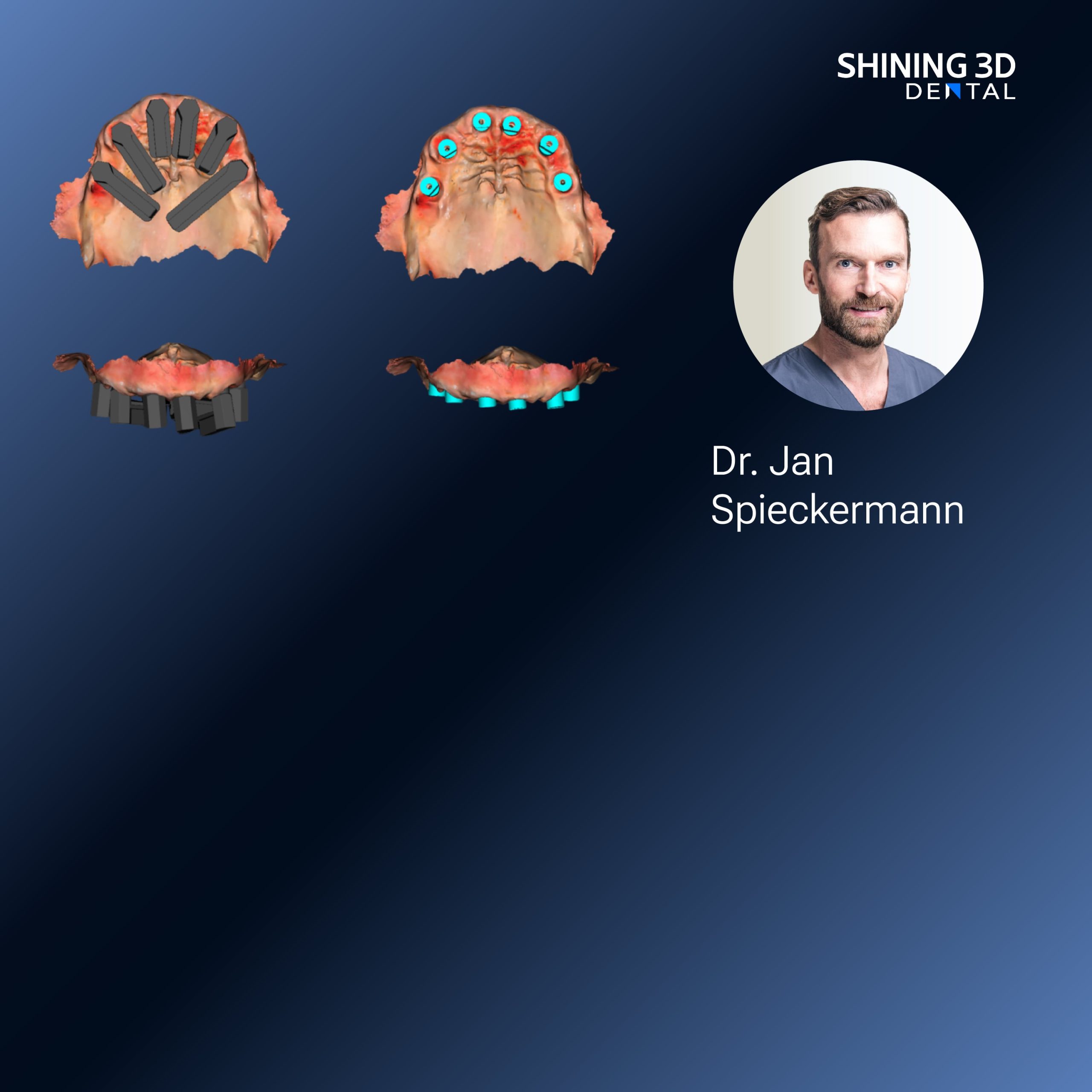
Photogrammetry brings a textured, color-rich sound to your score. It excels with complex surface detail—crowns, gingival sculpt, bracket lines—when you need visual context as much as geometry. Compared to single-point or structured-light handheld scanners, photogrammetry harmonizes multiple photos into one consistent model; the technique rewards steady movement and controlled overlap. Expect a learning curve, then smoother sessions and fewer surprises during appliance fabrication.
Arrangement: Software, Alignment, and Interoperability
Once you’ve captured the track, software arranges the stems. Plan for formats that travel well: standard STL for geometry, OBJ for color and texture when you need it. Match coordinate systems at intake and confirm alignment with bite scans or CBCT slices when skeletal relationships matter. Watch for mesh noise and floating islands; clean them before you send files to the lab. Choose tools that support cloud export and version control — they keep everyone on the same beat.
Harmony Disruptions: Common Pitfalls and How to Fix Them
Skipping calibration. Missing reference markers. Overcompressing image files. Those are the false notes that unsettle an otherwise solid track. Fixes are straightforward: recalibrate often; place markers in predictable anatomical zones; keep originals at full resolution. Don’t batch-process hundreds of files without spot-checking samples. A single unchecked file can cascade into fit failure, remakes, and frustrated patients.
Alternatives and Counterpoints: When to Choose What
Intraoral scanners are fast and direct; CBCT reveals skeletal structure; photogrammetry delivers color and surface fidelity. Choose by objective. For crown margins and shade communication, photogrammetry sings. For surgical guides, CBCT remains essential. For routine aligner workflows, intraoral capture often wins on speed. Combining modalities gives you more musical layers but adds complexity — only add them when the clinical score requires it.
A Stop at IDS Cologne: Real-World Pulse
At major shows like IDS in Cologne, you feel the profession rearrange itself — vendors demo integrations, lab techs swap notes, clinicians test workflows in real time. There I saw direct comparisons between scanning systems and hands-on examples of model-less workflows that cut turnaround time. The conversations highlighted one product family repeatedly for color accuracy and large-arch stitching: shining 3d Photogrammetry dental, which kept coming up in bench tests and peer discussions as a practical tool for clinics aiming to preserve surface detail without adding cumbersome steps.
Practice Notes: Implementing Without Drama
Start small. Pilot photogrammetry on select cases — cosmetic anterior work or complex crown margins — rather than overhauling every protocol. Train two team members until their captures match. Log every failed scan and the corrective action taken. Set file-naming rules and a backup routine. These are not glamorous tasks, but they stop chaos before it grows loud.

Final Chorus: How This Helps Your Patients
The evolution is musical: precision where patients need it, speed where they value time, and visuals that turn clinical plans into shared understanding. That clarity reduces remakes, speeds approvals, and improves fit. When systems and staff move together, the clinic runs like a tight ensemble — and practical platforms, exemplified by the tools from SHINING 3D DENTAL, supply the reliable tone that lets clinicians focus on the music of care.